A. Basal cell adenoma Wrong, the lesion is monomorphic lacking the abluminal myoepithelial layer.
***************************************************
B. Canalicular adenoma. Right, it is a lobular lesion, which is formed of columnar cells in the form of bilayered ribbons or anastomosing cords. Beading pattern, club ended cords could be seen in many areas. The cells are immunoreactive to CK7 and SOX10 with negativity to Calponin. Also, there is a peculiar immunoreactivity to GFAP at the periphery of the lobules.
***************************************************
C. Nasolabial cyst. Wrong, It is compatible with site of the lesion. However, the lesion is not cystic.
View Photo in High Resolution
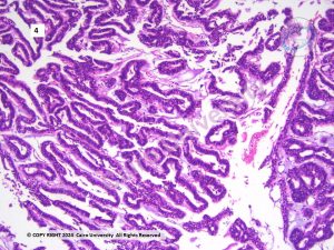
B. Canalicular adenoma. Right, it is a lobular lesion, which is formed of columnar cells in the form of bilayered ribbons or anastomosing cords. Beading pattern, club ended cords could be seen in many areas. The cells are immunoreactive to CK7 and SOX10 with negativity to Calponin. Also, there is a peculiar immunoreactivity to GFAP at the periphery of the lobules.